Exploring the Role of Adventitial Fibers in Cerebral Artery Biomechanics for the Subfailure Damage Regime
Abstract
The gradual recruitment of tortuous medial collagen fibers underlies the nonlinear behavior of arteries within the physiological loading regime, while adventitial collagen fibers are hypothesized to act primarily as a protective element during supraphysiological loading events, such as balloon angioplasty. We examined five human cerebral artery specimens using multiphoton microscopy to image adventitial collagen during progressive supraphysiological uniaxial testing to failure. Fiber angles, tortuosity, and sublayer location were quantified for 3467 fibers across the adventitial thickness. A mixed-effects regression model assessed the effects of strain, tortuosity, sublayer index, and their interactions on circumferential fiber alignment. Higher strain significantly decreased fiber angles (p < 0.001), indicating greater alignment, while higher tortuosity reduced alignment (p < 0.01) and diminished aligning effects of strain (p < 0.001). Sublayer position was also significant (p < 0.001), with adventitial fibers closer to the media being more aligned. With increasing stretch, adventitial fibers first lose tortuosity. At higher stretch (λ* ≈ 1.25 ± 0.09, σ* ≈ 0.92 ± 0.27 MPa), internal elastic lamina (IEL) tearing and adjacent medial fiber rupture occur, followed by circumferential adventitial fiber alignment. However, in this cohort (mean age 73.4 ± 7.9 years), realignment was observed after multiple transverse IEL and medial tears had occurred. These findings provide quantitative, layer- and sublayer-specific evidence of a two-stage adventitial response, highlighting the coupled influence of loading, tortuosity, and wall depth on fiber alignment, and underscoring the need for high-fidelity models that explicitly incorporate layer-specific fiber architecture and failure mechanisms.
Motivation
Cerebral arteries, including those of the Circle of Willis, normally carry modest circumferential wall loads. During interventions such as balloon angioplasty for cerebral vasospasm, stenting and endarterectomy, however, they can be stretched far beyond the physiological range, and unintended damage can lead to complications such as arterial dissection or in-stent restenosis. Most arterial mechanics research has focused on the physiological regime, so how the wall responds to supraphysiological loading, and how it begins to fail, is much less understood.
In the three-layered arterial wall, medial collagen is largely circumferential, while adventitial collagen has a wide spread of fiber angles. The adventitia is often treated as a passive connective sheath and is thought to act mainly as a protective layer at high loads, but this role has rarely been tested in human cerebral arteries. Earlier studies typically measured either the mechanics or the fiber microstructure, not both at once, and many relied on animal models whose anatomy and disease state differ from those of human brain arteries. Building on the method of Hill et al. for measuring medial collagen recruitment during loading, this study extends simultaneous mechanical testing and multiphoton imaging to the adventitia of the human middle cerebral artery, loaded progressively to failure.
Methodology
- Specimens: five human Circles of Willis obtained post-mortem through the University of Pittsburgh Brain Bank (2 males, 3 females; mean age 73.4 ± 7.9 years, range 61–82) and stored in HypoThermosol at 4 °C. Middle cerebral arteries (MCAs) were dissected within 24 h, and vessels with severe calcification or atherosclerotic plaque were excluded. Each MCA was opened longitudinally and a circumferentially oriented “dogbone” specimen was cut with a custom cutter to give smooth edges and consistent dimensions (central width 1.36 ± 0.02 mm, clamping width 3.54 ± 0.09 mm). All testing and imaging was finished within 36 h of harvest.
- Integrated uniaxial testing and multiphoton microscopy (UA-MPM): a custom uniaxial device under the microscope stretched each specimen circumferentially with a linear actuator at 20 μm/s while a 5 lb load cell recorded force. The tissue sat in a phosphate-buffered saline bath at room temperature, and a camera viewing the luminal side through a mirror tracked the internal elastic lamina (IEL). Cauchy stress was computed as force times stretch divided by the unloaded cross-sectional area, assuming incompressibility.
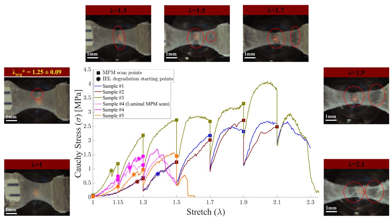
- Loading protocol: no cyclic preconditioning was applied, to avoid altering the native microstructure. Starting from an unloaded state (time-averaged preload of 0.01 N), specimens were held at stretch levels of 1.0, 1.3, 1.5, 1.7 and then in steps of 0.2 until complete failure, with an additional step at 1.15 for three specimens. Each hold lasted about 30 minutes while the specimen was imaged.
- Imaging: label-free second harmonic generation imaging of collagen from the abluminal (adventitial) side on a Nikon A1R MP HD system with a 25× water-immersion objective (NA 1.10) and an 820 nm laser. Each scan covered 500 μm × 500 μm with 0.5 μm in-plane resolution and 0.5 μm z-steps through the full adventitial thickness.
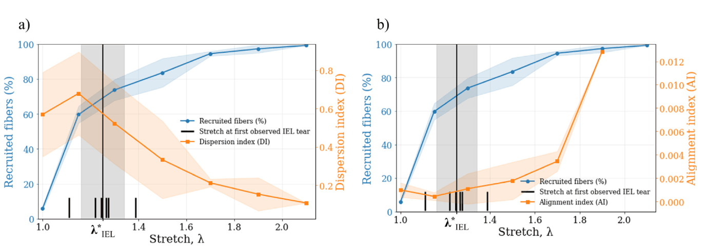
- Fiber analysis: at each stretch, three substacks of 10 slices (5 μm) were taken at fixed relative depths in the adventitia (outermost, middle and innermost, next to the media) and collapsed by maximum intensity projection. Between 35 and 55 fibers per substack were traced by hand in ImageJ by an operator who did not know the stretch level, for a total of 3467 fibers. Tortuosity was the ratio of a fiber’s arc length to its end-to-end distance, and a fiber was counted as recruited when its tortuosity was at most 1.02. Fiber angle came from a linear fit to the traced points and was weighted by fiber length, so the result does not depend on how a fiber was segmented. Stretch-specific dispersion and alignment indices were computed from Gaussian-peak fits of the angle distributions.
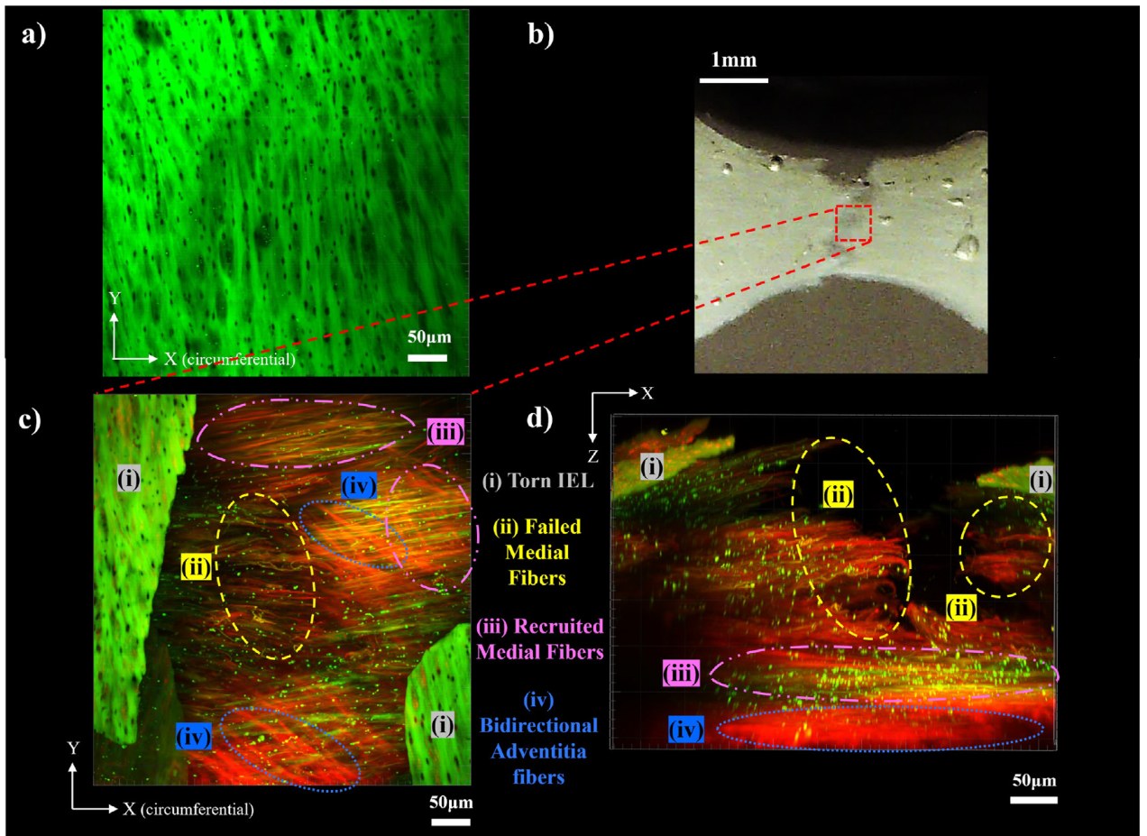
- Layer thickness and IEL imaging: media and adventitia thicknesses were measured on MPM cross sections of fresh tissue (at least 30 measurements per sample). A second specimen cut next to Sample 4 was mounted lumen-side up and imaged to follow the IEL and the medial fibers beneath it during loading.
- Statistics: a linear mixed-effects model (Python statsmodels) related each fiber’s absolute angle from the circumferential direction to strain, tortuosity, sublayer index (1 = outermost, 3 = innermost) and the interactions strain × tortuosity, strain × sublayer and tortuosity × sublayer. Strain and tortuosity were shifted to be zero in the unloaded, straight state and scaled by their standard deviations, and a random intercept per sample captured between-specimen variation.
Key Findings
- Wall geometry: the adventitia was 104 ± 30 μm thick and the media 216 ± 24 μm, giving an adventitia-to-media ratio of 0.48 ± 0.14. The adventitia is therefore a substantial part of the cerebral artery wall, not a thin sheath.
- IEL fails first: IEL tearing began at a stretch of 1.25 ± 0.09 and a Cauchy stress of 0.92 ± 0.27 MPa, far above the estimated physiological wall stress (second Piola–Kirchhoff stress of about 0.032 ± 0.005 MPa at a transmural pressure of 140 mmHg). More IEL tears appeared as loading continued, and all specimens failed in the narrow central region of the dogbone.
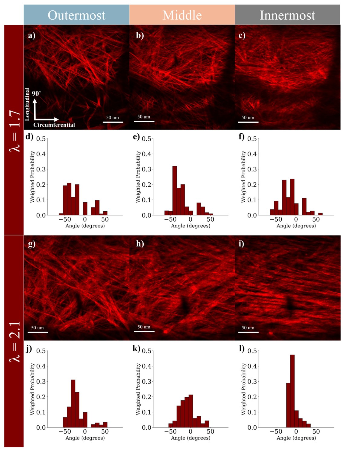
- Unloaded adventitia: before stretching, adventitial fibers were tortuous and bidirectional, with angle peaks at −61.4° and +58.9° from the circumferential direction (means −50.05° ± 21.82° and 48.92° ± 21.35°).
- Two-stage adventitial response: by a stretch of 1.3, the fibers had lost most of their tortuosity but kept a broad, bimodal angle distribution. Substantial reorientation toward the circumferential loading direction appeared by 1.7, and by 2.1 the distribution had collapsed to a single peak near 0°. In the representative sample, nearly 50% of innermost-layer fibers were oriented circumferentially at that stretch. Recruitment rose quickly at low stretch, while the drop in dispersion index and the rise in alignment index came later. The mean IEL tear-onset stretch fell between these two stages.
- Inner-wall damage under the lumen: in the specimen imaged from the lumen side, the IEL tore at a stretch of 1.3, with the tear running perpendicular to the loading direction. Medial fibers next to the tear also failed and appeared tortuous, while deeper medial fibers stayed straight and recruited and the adventitial fibers below remained intact and bidirectional. Medial fibers were already nearly straight and circumferential before loading, which the authors attribute to the donors’ advanced age, and almost all were recruited by a stretch of 1.3.
- Mixed-effects regression: absolute fiber angle decreased with strain (coefficient −9.18°, 95% CI −10.87 to −7.48, p < 0.001) and with tortuosity (−2.45°, p < 0.01), from an intercept of 54.22°. The positive strain × tortuosity interaction (3.22°, p < 0.001) means that stretch aligns highly tortuous fibers less effectively. Fibers deeper in the adventitia, closer to the media, were more circumferential (−4.11° per sublayer, p < 0.001), and this depth effect did not depend on strain or tortuosity (interaction coefficients 0.15 and 0.41, not significant). The fixed effects explained 19.2% of the variance (marginal R²), rising to 25.1% with the specimen random effect.
- Heterogeneity and kinematics: in Sample 3, the outermost layer kept a bimodal distribution right up to failure. Some axially oriented fibers became more tortuous at high circumferential stretch, which the authors explain by lateral contraction under circumferential loading. An affine deformation model predicts the same overall trend toward circumferential alignment, but the measured distributions became sharper at high stretch than affine kinematics predicts, so the collagen reorientation is not purely affine.
Conclusions
In these older human middle cerebral arteries, the adventitia behaves like a reactive “safety net”. The IEL tears first, at stresses well above physiological levels, and the medial fibers next to the tears fail with it. The adventitial collagen, by contrast, first straightens and then reorients toward the loading direction, holding the wall together until much higher stretch. Fiber alignment depends jointly on strain, tortuosity and depth within the adventitia, with inner adventitial fibers more circumferential and closer in architecture to the media. The authors conclude that high-fidelity constitutive models of cerebral arteries need failure criteria for the IEL and the elastin-rich matrix, not only for fiber rupture, as well as layer- and sublayer-specific fiber architecture. Such models are relevant to procedures that deliberately overstretch vessels, such as balloon angioplasty.
The authors note several limitations. The specimens were opened, passive and tested in uniaxial tension at room temperature, so the results do not reproduce in vivo pressurization, biaxial loading, residual stress or smooth muscle tone. However, extra imaging showed no significant change in baseline adventitial fiber alignment after the vessels were opened and flattened. The donors were older, so the results apply most directly to aged arteries. Stress relaxation during the roughly 30-minute imaging holds was not analyzed, and the IEL could be imaged only from the lumen side, in one additional specimen. Future work includes studying younger donors, viscoelastic effects and IEL failure in more depth.
Research Impact
Citations
Available on Google Scholar
Field
Cerebrovascular Biomechanics and Arterial Wall Damage
Approach
Integrated Uniaxial Testing and Multiphoton Microscopy with Mixed-Effects Regression of Fiber Orientation